VeLight: A 3D Virtual Reality Tool for CT-based Anatomy Teaching and Training
Abstract:
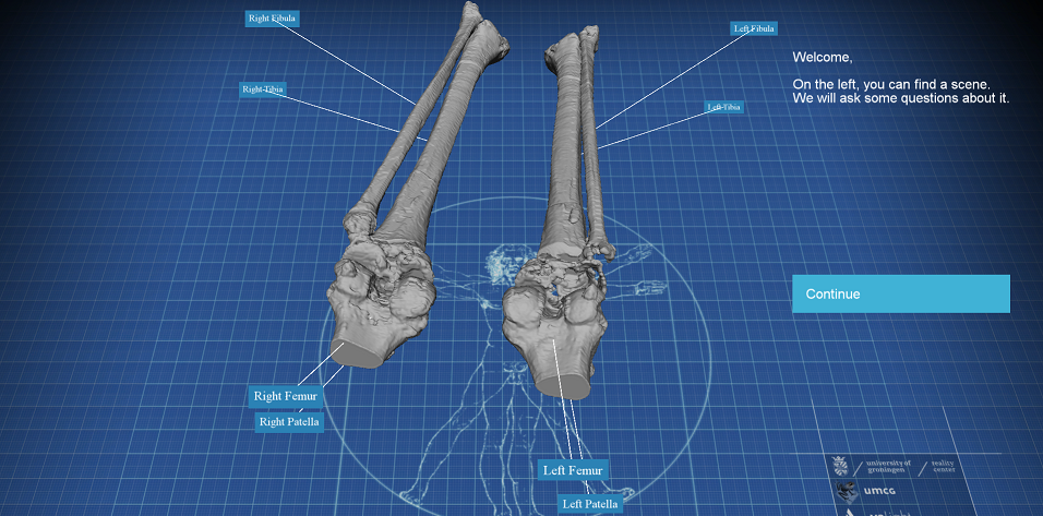
Anatomy is considered to be one of the most essential courses for medical students to develop their clinical skills (Fig. 1). A solid understanding includes, for instance, how the human skeleton is built up, the spatial relationships between body parts, the features of different organs, etc. Traditionally, anatomy is taught either by dissecting human cadavers or using text books. Dissection is considered as the most powerful means to presenting and learning the human body since it gives a direct observation and interaction with the human bodies. However, there are a few disadvantages of this method. First of all, preserving human bodies and maintaining dissection rooms in accordance with the European laws is extremely costly (Turney 2007). Besides, due to the limited space around a dissection table, only a small group of students can learn with one cadaver. However, the limited number of available cadavers does not allow a large number of dissections, which limits the opportunities for students to get sufficient anatomy education. Finally, the limitations of the regulations and number of cadavers does not enable self-directed learning, which is identified as an essential skill for medical graduates (Ramnarayan and Hande 2005). Therefore, most teaching of anatomy is still performed from books and online materials that are usually easy to reach and cheap. Another benefit is that all teaching materials in available books and other supporting materials can be found from given references or other online resources, which makes self-directed learning possible. So this method is easy for teachers to organize courses and teaching materials and for students to systematically acquire the required knowledge. However, most of the traditional text books focus on 2D images which is not sufficient to replace human dissection. 3D perception, compared to 2D, provides a better view of depth information of visualized data, which is very important in understanding the spatial location and relationships of human bodies and their composition (Tam et al. 2009; Drake et al. 2010). Another disadvantage is the fact that books show mostly only a specific, and typically a perfect, example of the human body. Therefore, it is not possible to learn from real use cases. Although these are sometimes enhanced with 3D images, for instance, the existing 3D augmented reality books (Wu et al. 2013) can pop up a 3D virtual object on top of the presented material, the interactive 3D experience for students is very limited.
Main reference:
Yu, L., Ouwerling, J., Svetachov, P. et al. VeLight: A 3D virtual reality tool for CT-based anatomy teaching and training. J Vis 25, 293–306 (2022). https://doi.org/10.1007/s12650-021-00790-y
BibTeX:
@article{yu2022velight,
  title={VeLight: A 3D virtual reality tool for CT-based anatomy teaching and training},
  author={Yu, Lingyun and Ouwerling, Joost and Svetachov, Pjotr and van Hoesel, Frans HJ and van Ooijen, Peter MA and Kosinka, Jiri},
  journal={Journal of Visualization},
  volume={25},
  number={2},
  pages={293--306},
  year={2022},
  publisher={Springer}
}